Skip to content

It's Not Just Fat.

It's Likely Lipedema.

Lipedema is a progressive medical condition affecting 1 in 9 women, yet few hear about it, even from their own doctor. 

It's not your fault, it's a real disease, Lipedema is treatable and is often covered by insurance.

Lipedema Stage 1
Lipedema Stage 2
Lipedema Stage 3
Lipedema Stage 4

That won't fix this.

Lipedema is a chronic condition that causes fat to build up in an unusual way on the legs, hips, upper buttocks, and often the arms. The tissue can feel tender or bumpy, bruise easily, and feel heavy or painful.

What sets it apart is that lipedema fat rarely responds to diet and exercise. You can lose weight from your upper body while your legs barely change. That isn't a lack of willpower. It's how the disease behaves.

It is an inherited genetic condition that often starts or gets worse at puberty, during pregnancy, or around menopause, and it frequently runs in families.

Lipedema is often mistaken for ordinary weight gain, even by doctors. That's why so many women spend years blaming themselves for something they didn't cause.

Does this sound familiar? (two or more would be a reason to speak to a specialist)

  • Your lower body is out of proportion with your upper body
  • Diet and exercise haven't changed your legs
  • Your ankles look like they have a "cuff" just above the foot
  • Your legs ache, feel heavy, or bruise easily
  • Your mother, sister, or aunt has the same shape

If you checked two or more, it's worth talking to a specialist.

Getting a diagnosis is the first step. From there, care is built around you:

  • Conservative care: compression, lymphatic therapy, and nutrition and lifestyle support to help manage symptoms
  • Lipedema reduction surgery: specialized techniques that remove diseased fat tissue while protecting the lymphatic system, for patients who are good candidates

You may not need surgery, and not everyone is a candidate. A specialist evaluation is the first step to starting your healing journey.
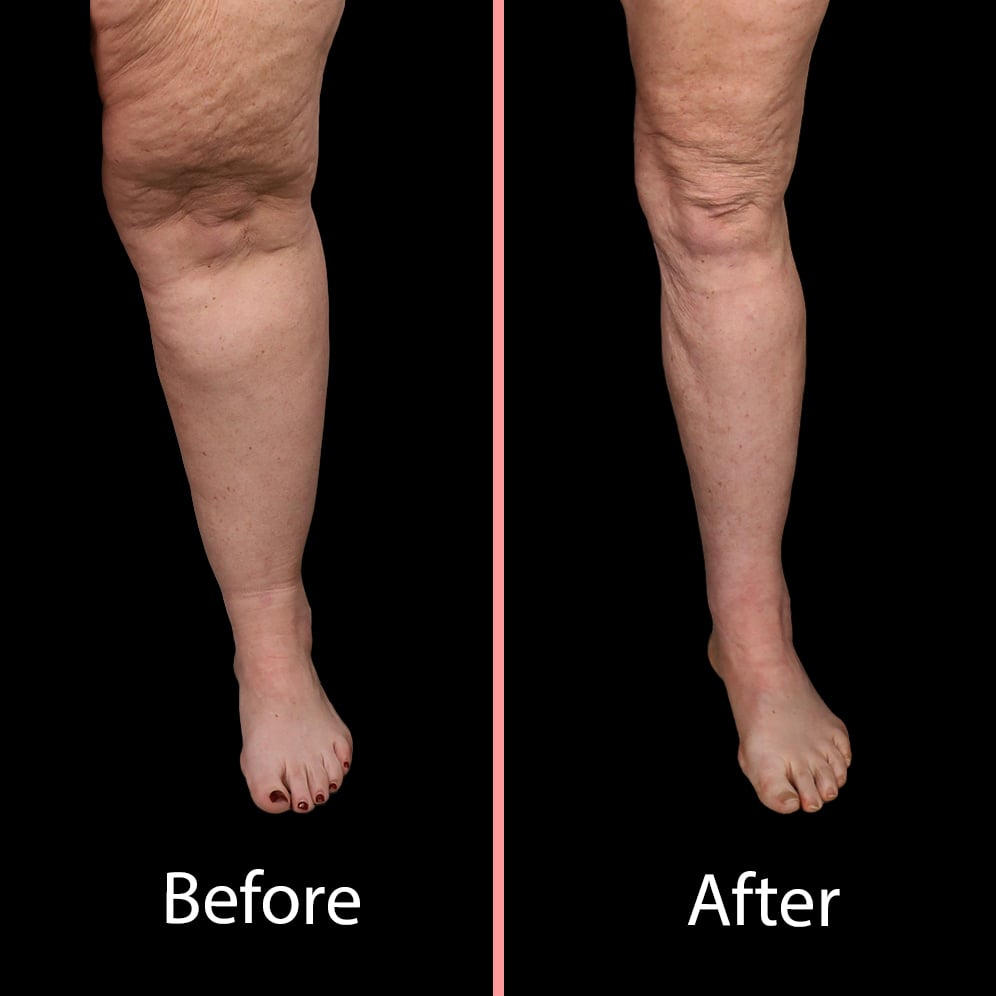
Lipedema before and after advanced lipedema treatment surgery with the roxbury institute

At The Roxbury Institute, lipedema isn't something we treat on the side. It's a core specialty, supported by research and a dedicated program: Advanced Lipedema TreatmentTM.
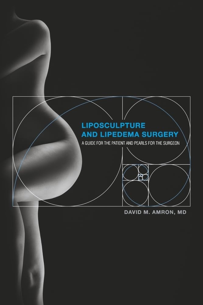

  • Led by Dr David Amron, a board-certified dermatologic surgeon with more than 25 years of experience and over 12,000 procedures, and author of Liposculpture and Lipedema Surgery
  • Diagnosis, conservative care, surgical intervention, lifelong disease management, and research all in one institute
  • Locations in Los Angeles CA, Salt Lake City UT, and Tucson AZ
  • Liposculpture And Lipedema Surgery: A Guide for the Patient and Pearls for the Surgeon by Dr. David Amron Book Cover Photo
  • The Roxbury Institute - Los Angeles Lipedema Surgery Center for Advanced Lipedema Treatment
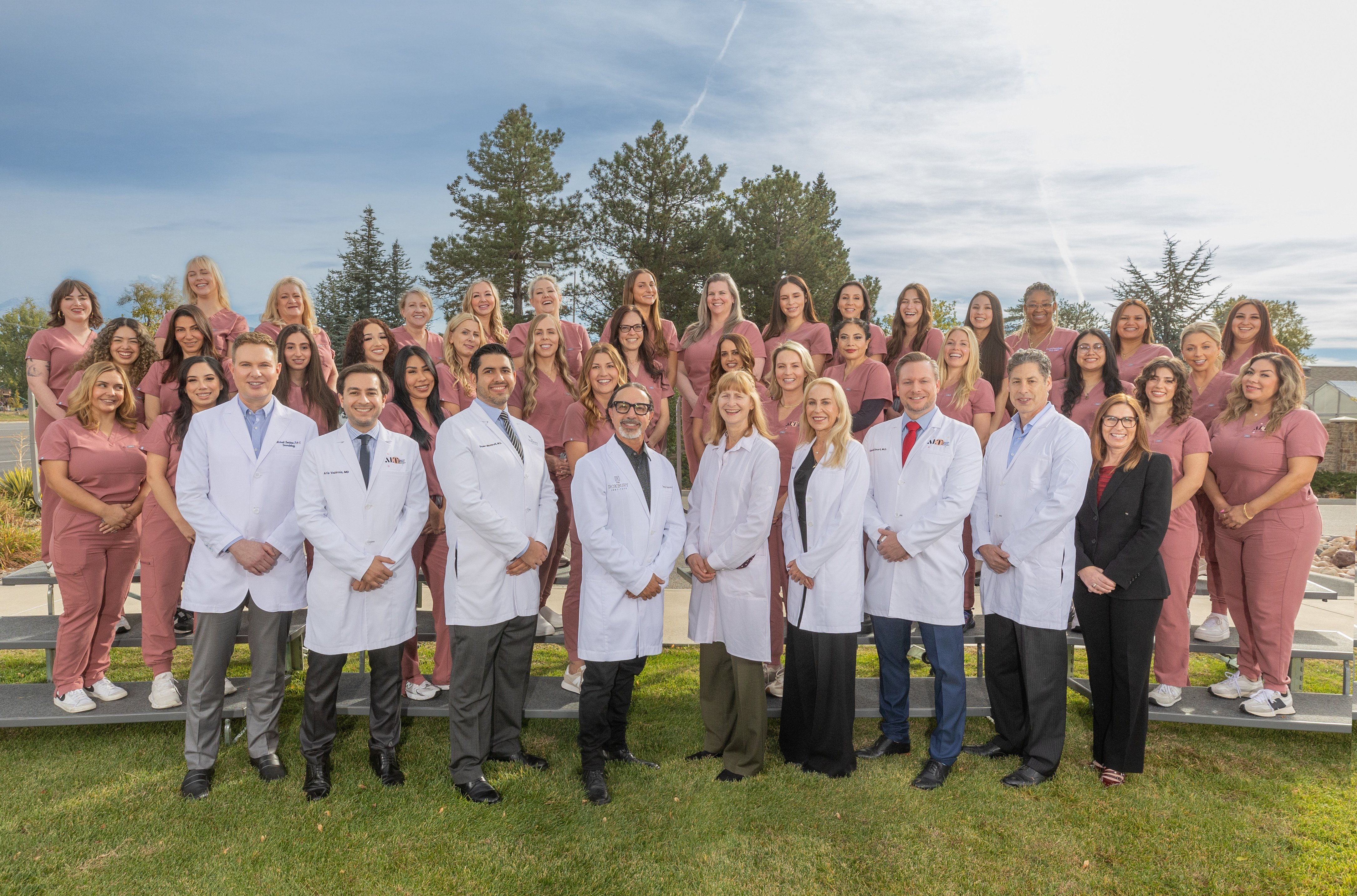
  • The Roxbury Institute Clinical Team Photo
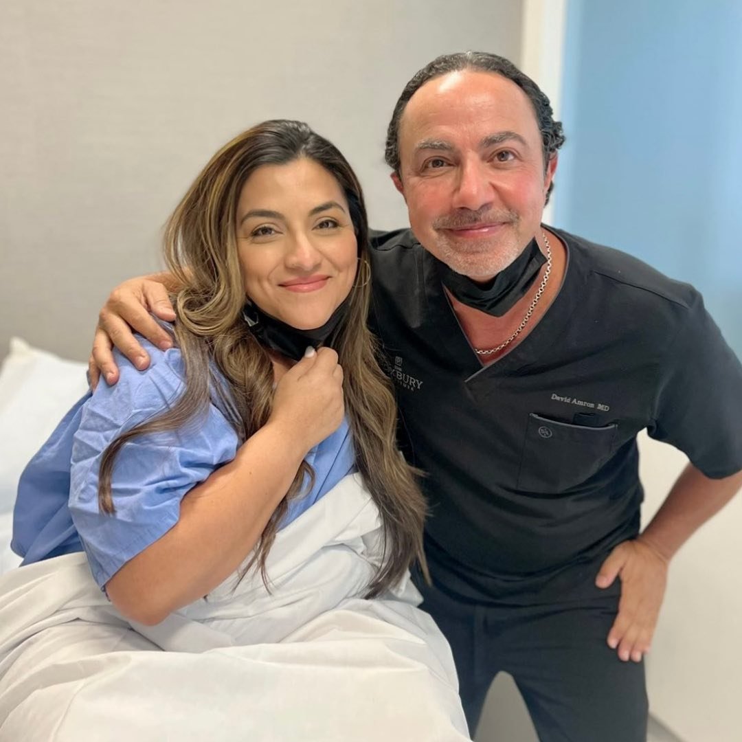
  • Dr David Amron with arm around patient

The first step in getting ahead of Lipedema is getting a diagnosis.

Submit a Contact Form Today.

Tell us a little bit about you so we can tell you about Lipedema and options you have.

We'll Connect.

Our Advanced Lipedema Treatment Team at The Roxbury Institute has information, consultation appointments available, experience with many insurers to help with surgery, and much more to discuss.

You Get Answers.

Lipedema is a lot, but it's not something you have to carry with you forever. We'll help you get answers to your burning questions, and options for your care. Including a diagnosis.

1. What is Lipedema, and how does it differ from obesity and lymphedema?

Lipedema is a chronic, progressive, disproportionate fat storage disease that primarily affects women. It is estimated to affect up to 11% of the female population—translating to roughly 17 million women in the United States and 370 million worldwide. Despite its high prevalence, Lipedema is widely omitted from standard medical school curricula. Consequently, primary care physicians and specialists frequently misdiagnose the condition as general obesity, lymphedema, or elephantiasis, leaving patients without appropriate care for decades.To provide a more medically precise description of the underlying pathology, Dr. David Amron proposed the diagnostic term Lipofibroedema. Breaking down the tripartite nature of this term reveals the true biological complexity of the disease:

  • Lipo: The abnormal, pathologic overgrowth and duplication of subcutaneous adipose (fat) tissue.
  • Fibro: The deposition of fibrotic scar tissue, chronic inflammation, and severe fascial tethering within the fat layer.
  • Edema: The chronic stagnation and accumulation of lymphatic fluid resulting from impaired vessel permeability.Lipedema differs fundamentally from general obesity and primary/secondary lymphedema in three key ways:
  • Symmetrical, Bilateral, and Circumferential Distribution: Lipedema fat accumulates symmetrically and bilaterally and wraps circumferentially around the entire extremity (arms or legs), destroying natural anatomical definition and creating heavy, "column-like" limbs.
  • Metabolic Resistance to Standard Weight Loss: Unlike non-Lipedema subcutaneous fat associated with lifestyle-related obesity which involves more normal fat tissue, Lipedema tissue is metabolically pathologic. It is less responsive to typical weight loss strategies like dieting, weight loss surgery, exercise, and GLP-1 Agonists.
  • Sparing of Hands and Feet: A hallmark diagnostic differentiator between Lipedema and lymphedema is that Lipedema tissue strictly spares the feet and hands, ending abruptly in a distinct tissue "cuff" at the ankles or wrists. Primary or secondary lymphedema, by contrast, involves impaired lymphatic transport that causes fluid swelling directly extending into the dorsum of the feet and hands.
2. What are the signs, stages, and types of Lipedema?

The physical signs of lipedema include a symmetrical, "column-like" fat pattern encircling the entire leg or arm while typically sparing the hands and feet, resulting in a disproportionate shape, with a narrow waist and larger hips, thighs, ankles, arms. This is often accompanied by sensitivity or pain to pressure, easy bruising, persistent swelling, and flexible or hypermobile joints. 

Lipedema is clinically evaluated and categorized using two main systems: Stages, which evaluate disease severity and structural tissue changes, and Types, which define the anatomical distribution of diseased tissue across the body.

Lipedema Stages

Stage 1

"Mild circumferential fullness; skin surface remains soft and smooth, though underlying microscopic tissue changes are present."

Stage 2

"Moderate circumferential fullness; skin surface irregularities develop (mattress-like texture) alongside palpable, walnut-sized subcutaneous fat nodules."

Stage 3

"Advanced circumferential fullness; large, overhanging lobules of fat deform the limbs, accompanied by marked tissue hardening (induration) due to progressive fibrosis."

Stage 4

"Very advanced circumferential fullness with massive tissue lobules; severe fibrotic hardening and chronic lymphatic vessel collapse lead to secondary lymphedema, a combined state known as Lipolymphedema ."

Lipedema Types
  • Type I: Diseased fat is localized to the pelvic girdle, involving the upper buttocks, hips, and lower abdomen.
  • Type II: Diseased fat extends from the pelvic girdle down through the thighs circumferentially, ending at the knees.
  • Type III: Diseased fat extends circumferentially from the pelvis all the way down to the ankles. This represents the most common clinical distribution pattern.
  • Type IV: Diseased fat involves the upper arms circumferentially, with frequent extension down the forearms to the wrists.
  • Type V: Diseased fat is isolated circumferentially to the lower legs, extending strictly from below the knees to the ankles.Regardless of anatomical type, Lipedema characteristically strips affected limbs of their normal visual contours. In the lower extremities, circumferential fullness obliterates the visible line of the tibia (shinbone) and hides the contours of the ankle bones. In the upper extremities, fat wraps uniformly around the top, sides, and undersides of the arm, pulling the tissue downward and causing the upper arm to lose its structural shape.
3. What causes Lipedema, and what is its underlying pathophysiology?

Lipedema is a genetically inherited, systemic connective tissue disorder that follows an autosomal dominant inheritance pattern. The gene can be inherited with equal frequency from either the maternal or paternal lineage; however, while men carry the genetic trait as frequently as women, the disease is expressed almost exclusively in females.Systemic manifestation and rapid acceleration of the disease are tied to major hormonal shifts. Specific hormonal trigger points include puberty, pregnancy, menopause, or post-OB-GYN surgical interventions , which induce hormonal shifts that accelerate fluid leakiness across compromised capillary networks.

Pathophysiology: The Vicious Cycle

Lipedema is not an eating disorder or a failure of willpower. It is a progressive disease driven by a self-perpetuating cellular feedback loop:

  1. Leaky Lymphatics (Lymphorrhea): Inherited connective tissue laxity compromises the structural integrity of subcutaneous lymphatic vessel walls, causing fluid to leak continuously into surrounding tissues.
  2. Cell Transformation & Hypertrophy: Fluid accumulation signals subcutaneous stem cells to differentiate into preadipocytes, which rapidly duplicate and expand (fat cell hypertrophy).
  3. Hydropic Degeneration: Fat cells duplicate so rapidly that they outpace their capillary blood supply, undergoing cell death via hydropic degeneration.
  4. Chronic Inflammation & Macrophage Infiltration: Macrophages infiltrate the subcutaneous layer to clear cellular debris from dying fat cells, triggering chronic tissue inflammation and laying down fibrotic scar tissue.
  5. Tissue Congestion & Hydrostatic Feedback: Accumulated fibrotic scar tissue hardens the subcutaneous plane, increasing tissue hydrostatic pressure and physical congestion. This pressure further crushes delicate lymphatic channels, causing additional fluid leakage and perpetuating the vicious cycle.
Co-Occurring Conditions

Approximately 80% to 85% of Lipedema patients present with co-occurring Ehlers-Danlos syndrome (EDS) , a genetic collagen disorder characterized by joint hypermobility, systemic tissue fragility, and hyper-flexible skin. This high rate of co-occurrence reinforces that Lipedema is fundamentally rooted in systemic collagen and connective tissue defects rather than isolated adipocyte dysfunction.

4. What are the primary symptoms associated with Lipedema?

Lipedema manifests through a combination of physical, systemic, and emotional symptoms that impact daily living and functional mobility.

Physical Symptoms
  • Pain & Pressure Sensitivity: Sensations range from a persistent, deep dull ache to sharp, burning pain. Affected limbs are acutely sensitive to touch, light pressure, or minor impact (such as a pet resting on the lap or a light clinical examination).
  • Fluid Swelling (Edema): Fluctuating tissue swelling that worsens with prolonged standing, high environmental heat, physical stress, or air travel.
  • Capillary Fragility & Easy Bruising: Microvascular fragility causes pronounced, spontaneous bruising from minimal contact or without remembered trauma.
  • Joint Hypermobility & Loose Skin: Hypermobile joints (particularly in the knees, hips, and fingers) and pliable, easily stretched skin due to underlying collagen defects.
  • Impaired Proprioception & Balance: Tissue swelling and deep fibrosis compress peripheral sensory nerves, impairing kinesthetic awareness, spatial orientation, gait, and overall balance.
Misdiagnosed Conditions

Because fibrotic tissue accumulation compresses distal nerves and alters circulation, Lipedema pain is frequently misdiagnosed as:

  • Plantar fasciitis
  • Fibromyalgia
  • Reflex sympathetic dystrophy (RSD / CRPS)
  • Peripheral neuropathy
  • Venous insufficiency
Emotional Impact

The emotional toll of Lipedema is profound. Most patients endure decades of medical gaslighting, where their symptoms are dismissed as simple overeating or lack of exercise discipline. This chronic invalidation often produces severe body dysmorphia, intense guilt over failed weight-loss attempts, deep depression, and overwhelming anxiety regarding future loss of mobility.

5. Why don't traditional weight loss, diets, or GLP-1 medications cure Lipedema?

Standard weight loss interventions fail to eliminate Lipedema because Lipedema tissue consists of metabolically locked, pathologic fat encapsulated in dense, fibrotic scar tissue.

  • Caloric Restriction, Exercise, and Bariatric Surgery: Standard weight loss methods burn normal non-Lipedema subcutaneous fat. When a patient with Lipedema undergoes caloric restriction, extreme exercise, or bariatric surgery, they lose weight almost exclusively in non-affected zones (such as the face, neck, and upper torso). The fibrotic Lipedema fat in the extremities remains largely unchanged, making the patient's body disproportion even more pronounced. Bariatric surgery should not be utilized as a primary therapy for Lipedema.
  • GLP-1 Receptor Agonists (e.g., Ozempic, Wegovy, Mounjaro, Rybelsus, Zepbound): These medications promote weight loss by delaying gastric emptying and suppressing central appetite. While some Lipedema patients report a secondary reduction in generalized systemic pain or systemic inflammation, GLP-1 agonists alone generally have a minimal impact on the break down of, and do not remove dense fibrotic Lipedema tissue .
  • Risk of Delaying Disease-Specific Treatment: Relying on weight loss drugs or aggressive dieting as a primary strategy creates a false sense of therapeutic progress. Meanwhile, underlying lymphatic leakage and fibrotic deposition proceed unchecked, increasing the risk of disease progression into Stage 4 Lipolymphedema. Patients must be protected against predatory commercial clinic behaviors and piecemeal "à la carte" pricing models that promise weight loss while failing to address the underlying structural disease.
6. What non-surgical treatments and nutrition strategies help manage Lipedema?

Non-surgical, conservative modalities are essential for managing daily fluid swelling, slowing disease progression, and reducing tissue inflammation.

Compression Therapy

Compression is vital for managing fluid dynamics and supporting compromised connective tissue:

  • Garment Selection (Flat-Knit vs. Circular-Knit): Custom flat-knit garments are mandatory for Stage 2, 3, and 4 disease. Flat-knit fabric is thick, stiff, and woven flat, providing a firm fabric wall that spans deep skin folds and lobules without pinching or creating tourniquet effects. Conversely, circular-knit garments are elastic and seamless, making them suitable only for mild, non-lobular Stage 1 tissue or light daily support.
  • Bandaging & Wraps: Multilayered, short-stretch compression bandaging or adjustable velcro compression garments (such as FarrowWraps) provide high resting pressure to reduce limb volume during acute swelling flares.
  • Pneumatic Compression Pumps: Advanced pneumatic devices (such as the Flexitouch or Lymphopress Optimal Plus) utilize multi-chambered sequential compression sleeves to gently drive trapped lymphatic fluid out of congested extremities toward functional lymph drainage basins in the torso.
Physical Therapies
  • Manual Lymphatic Drainage (MLD): A specialized, light-touch manual therapy designed to stimulate lymphatic vasomotion and reroute fluid around congested subdermal pathways.
  • Aqua Therapy: Water-based movement utilizes natural hydrostatic pressure to promote fluid clearance while buoyancy protects hypermobile, painful joints.
  • Vibrational Therapy: Low-frequency whole-body vibration platforms help stimulate lymphatic circulation and loosen congested fluid.
  • Quadrivas / Subcutaneous Adipose Tissue (SAT) Therapy: A specialized deep-tissue manual therapy designed to systematically manipulate the subcutaneous adipose tissue plane, break down superficial fascial adhesions, enhance tissue mobility, and improve functional limb volume.
Nutrition & Supplements
  • Anti-Inflammatory Dietary Frameworks: Patients benefit from nutritional frameworks that minimize systemic inflammation and fluid retention, such as the Rare Adipose Disorder (RAD) diet, whole-food plant-based diets, low-carbohydrate protocols, or ketogenic diets. Strict exclusions include refined grains, gluten, added sugars, chemically modified oils, and high-sodium processed foods.
  • Targeted Supplements: Recommended anti-inflammatory and vascular-support supplements include Diosmin, Micronized Purified Flavonoid Fraction (MPFF), Vitamin D3, Omega-3 fatty acids, Turmeric (Curcumin), Quercetin, Bioflavonoids, Vitamin C, Calcium, Magnesium, Green Tea extract, L-Carnitine, Selenium, Chromium, Butcher’s Broom, Dandelion Root, L-Arginine, Pycnogenol, Alpha-Lipoic Acid, and Ashwagandha.
7. How does Lipedema liposuction differ from cosmetic liposuction?

Liposuction for Lipedema is a specialized surgical procedure. It requires medical protocols, safety parameters, and surgical techniques that differ fundamentally from elective cosmetic body contouring.

  • Core Therapeutic Objective: Cosmetic liposuction only focuses on aesthetic contouring and localized fat removal in healthy tissue. Lipedema liposuction is a disease-directed functional surgery designed to clear pathologic fat, decompress trapped lymphatic channels, remove painful fibrotic scar tissue, and halt disease progression while maximizing three-dimensional cosmetic restoration.
  • Anatomical Scope & Technical Execution: Lipedema surgery requires specialized, lymphatic-sparing protocols capable of extensively extracting deep and subdermal fibrotic tissue. It demands technical expertise in complex anatomical zones that cosmetic surgeons generally avoid, including the calves, ankles, forearms, and wrists.
  • Circumferential Treatment vs. Spot Sculpting: Cosmetic liposuction often spot-treats isolated fat deposits. Lipedema surgery requires treating full anatomical units circumferentially to remove all active, diseased tissue across the entire region.
  • Perioperative Risk Management: Lipedema patients possess fragile capillary beds, high susceptibility to post-surgical bruising, and an elevated baseline risk for Deep Vein Thrombosis (DVT) due to chronic tissue congestion. Consequently, Lipedema protocols require strict perioperative medical management, including routine prophylactic anticoagulation therapy and IV hydtration.
8. What is the 360° circumferential surgical approach and how is it performed?
Comparing Surgical Approaches
  1. Spot Treating / Debulking: Focuses exclusively on localized "hotspots" (such as the medial knee bulge). While this offers short-term localized pain relief, it leaves active diseased tissue in adjacent untreated zones. This frequently causes rapid symptom recurrence, ongoing limb disproportion, and early revision surgery.
  2. Strip Approach: Treats one longitudinal strip of a limb at a time (e.g., inner thigh only, or anterior leg only) across separate surgical sessions. This approach is reserved for the 2% to 3% of advanced Stage 3/4 or Lipolymphedema patients whose tissue is too congested, vascularly compromised, or unhealthy to safely tolerate full circumferential treatment in one session.
  3. 360° Circumferential Approach: Treats an entire anatomical "cosmetic unit" (e.g., the complete thigh unit from groin to knee, or the entire lower leg unit from knee to ankle) three-dimensionally in a single operation. Executed in over 95% of Lipedema cases, this approach clears the disease completely from the affected unit, enables uniform skin retraction, and yields balanced functional and aesthetic outcomes.
The 5-Step ALT Liposculpture Process at The Roxbury Institute
  • Step 1: Tumescent Anesthesia (Hydrodissection): Using Water-Assisted Liposuction (Body-Jet) or a specialized Klein pump, large volumes of dilute lidocaine, epinephrine, and sodium bicarbonate are gently infiltrated into the subcutaneous fat layer. Epinephrine and hydrostatic pressure induces microvascular vasoconstriction to create a virtually bloodless operating field, while the fluid hydrodissects (forces open) dense tissue planes. To maintain strict patient safety, the surgeon adheres to a safe maximum lidocaine threshold of 55 mg/kg of body weight (frequently utilizing half-strength concentrations to safely treat large surface areas).
  • Step 2: Debulking (Vibrosuction): Utilizing Power-Assisted Liposuction (PAL) with high-speed mechanical vibration—frequently with the suction temporarily disengaged ("vibrosuction")—the surgeon safely releases dense fibrotic scar tissue and debulks the deep pathologic fat layer without tearing structural tissues.
  • Step 3: Emulsification (VASER): High-frequency ultrasonic energy is delivered directly into the debulked layer. VASER sound waves liquefy remaining fat cells and melt fine micro-fibrosis under the dermis. This creates an emulsified, oily matrix that protects overlying skin, transects fibrous cellulite bands, and allows smooth, uniform skin retraction.
  • Step 4: Fine Sculpting in the "Safe Plane": Using small, specialized cannulas, the surgeon executes precise fine sculpting within the strictly defined "safe plane" —the subcutaneous fatty layer situated strictly between the overlying dermis and the underlying deep muscle fascia. Operating parallel to the fascial plane prevents catastrophic complications such as muscle trauma, major vessel perforation, or internal organ injury, while avoiding surface deformities like skin tethering or buttock drop (ptosis). Under twilight sedation, the patient periodically tightens their underlying muscle groups, providing a stable platform for precision sculpting.
  • Step 5: Skin Tightening: Controlled thermal energy devices utilizing radio-frequency (Quantum) or superheated helium plasma (Renuvion) are applied beneath the skin to contract the soft tissue matrix in areas with moderate laxity. (If severe, irreversible skin laxity is present, excisional procedures such as a brachioplasty, thighplasty, or abdominoplasty are staged concurrently or separately).
9. What should patients expect during postoperative recovery?
Immediate Post-Op (24–48 Hours)

Tiny incisions are lightly closed using a small dissolving suture without the placement of permanent surgical drains. For the first 12 to 36 hours, blood-tinged tumescent fluid will drain naturally from the incision points. Absorbent padding is secured beneath compression garments to manage fluid drainage. Patients are required to perform light walking immediately after surgery to promote venous return and prevent clot formation; however, they must be assisted by an adult at all times during the first 24 hours due to temporary fluid shifts. Fluid hydration (drinking a glass of water every hour) is mandatory.

Critical Safety Rule: Showering Protocol

For the first 4 days following surgery , patients must shower directly in their compression garments . Removing compression garments prematurely during this initial period releases tissue pressure suddenly, causing massive fluid shifts into the lower extremities. This can trigger severe postural hypotension, sudden drops in blood pressure, lightheadedness, and fainting. Showering in the garment preserves systemic hemodynamic stability until vascular tone adapts.

Medical Management & Anticoagulation

Because Lipedema patients carry an elevated risk for thromboembolic events, a mandatory prophylactic anticoagulation regimen—specifically daily subcutaneous Lovenox (enoxaparin) injections —is initiated the day after surgery and continued for 5 consecutive days to prevent Deep Vein Thrombosis (DVT).

Compression Garment Protocol

Compression garments are applied on the operating table immediately upon surgical completion. The mandatory garment protocol requires:

  • Month 1: Worn 24 hours a day continuously (except during controlled garment changes after Day 4).
  • Month 2: Reduced to 12 hours a day (typically during daytime hours), provided linear decongestion is progressing normally.
Decongestion & Healing Timeline

While most patients experience linear decongestion and immediate pain relief, a subset of patients act as "delayed decongesters." These individuals experience a temporary increase in localized swelling and tissue congestion during the initial weeks following surgery as their compromised lymphatic system reorganizes. Initial structural changes are visible within 4 weeks; however, final tissue decongestion, skin retraction, and complete visual results typically take between 6 to 18 months.

10. Does Lipedema return after liposuction surgery, and how is it prevented long-term?

When Lipedema fat and its surrounding fibrotic scar tissue are properly removed using specialized, lymphatic-sparing techniques, the disease rarely recurs to any significant degree in those treated anatomical zones . Clinical tracking spanning over two decades demonstrates long-term disease stability following proper surgical extraction.

Long-Term Prevention & Maintenance Strategies
  1. Adhere to Anti-Inflammatory Nutrition: Maintaining an anti-inflammatory dietary framework (such as the RAD diet, ketogenic, or whole-food plant-based diet) long-term prevents systemic inflammation from re-triggering capillary leakiness in unoperated tissue planes.
  2. Proactive Trigger Management: Patients must identify and protect against personal swelling triggers. Wearing compression garments during high-risk events—such as long-haul air travel, extended automobile travel, high environmental heat, or intense exercise—prevents secondary lymphatic fluid accumulation.
  3. Lifelong Care for Aggressive Swellers: A small subset of patients ( <1% ) presenting with extreme baseline swelling or advanced Stage 4 Lipolymphedema remain at elevated risk for ongoing fluid retention post-operatively. These patients require lifelong maintenance therapy, including custom compression garment wear, periodic MLD, or pneumatic compression pump usage.Surgical treatment serves as a physical reset. By clearing diseased fat and removing dense fibrotic obstruction, surgery restores health to the subcutaneous tissue plane, providing patients with a functional baseline that can be successfully maintained for life.
  4. Some patients may benefit from micro or low dose GLP-1 for long term inflammation benefits.
  5. Specific supplements have been shown to help reduce inflammation, swelling, and optimize metabolism as part of a long term lifestyle strategy.
  6. Exercise. Motion. Movement.